Exercise Preserved Mitochondrial Energy Production Into the 90s. It Didn’t Protect the Circuit Breaker.
Prefer to listen? Hit play for a conversational, audio‑style summary of this article’s key points.
Much of what we call "mitochondrial aging" in muscle may be inactivity, not age. Older people move less, and muscle mitochondria respond to how much you move on a timescale of days, not decades. Nearly every study comparing young and old muscle has unknowingly compared active people to sedentary ones. This study broke that confound by profiling 139 men aged 20 to 93 and sorting them by physical activity, not just age, so it could ask what genuinely changes with age once movement is accounted for.
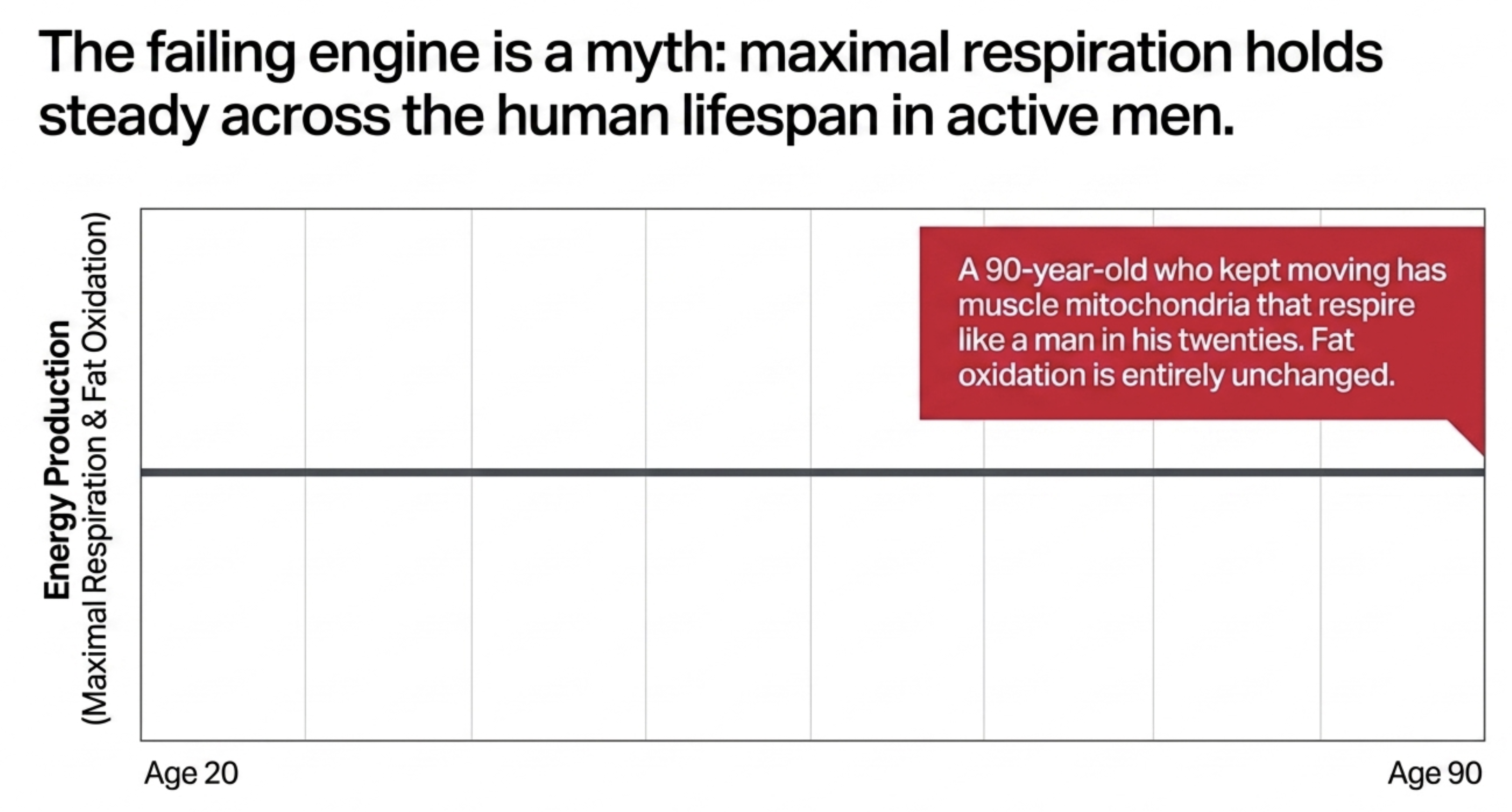
Mitochondrial energy production does not decline with age. In men who stayed active, maximal respiration held steady across the entire adult lifespan; a man in his nineties had mitochondria that respired like a man in his twenties. Where a decline appeared in inactive men, it tracked how little they moved, not how old they were. Exercise does not build better mitochondria, it builds more of them: when respiration was measured per unit of mitochondria, the differences between young and old, and between active and inactive, disappeared.
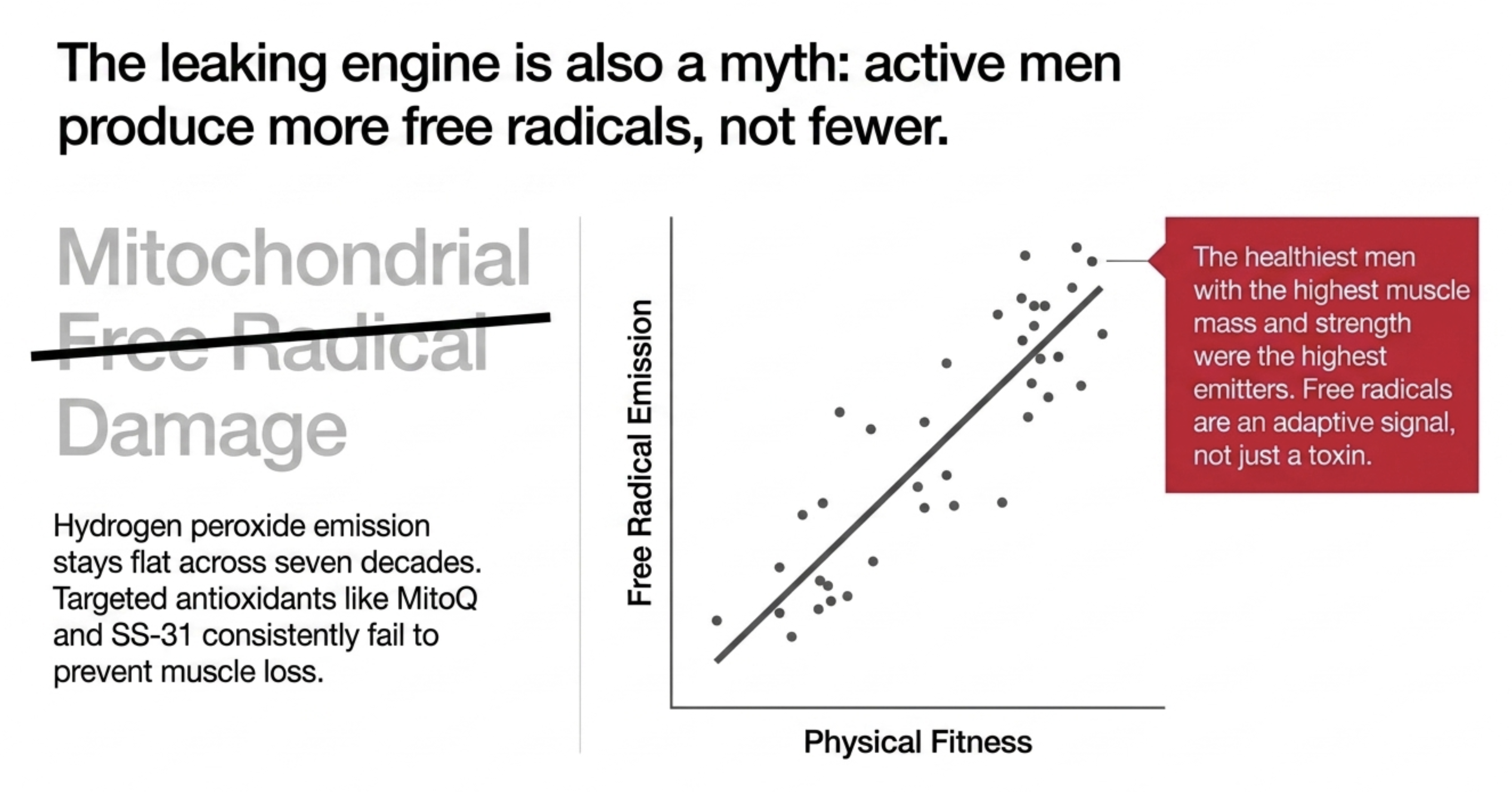
Oxidative "damage" does not rise with age either. Mitochondrial free-radical production was flat across seven decades, and, counterintuitively, active men produced more of it, not less, because free radicals are partly a signal the muscle uses to adapt to exercise. Critically, the men who produced the most showed no loss of muscle mass, strength, or function. The free-radical theory predicts the opposite, and it did not hold.
This undercuts the rationale for mitochondria-targeted antioxidant supplements. If aging muscle does not overproduce mitochondrial free radicals, there is nothing for these supplements to correct. Consistent with that, mitochondria-targeted antioxidants have repeatedly failed to prevent muscle loss in animal studies, and mice engineered for lifelong high oxidative stress do not develop accelerated muscle aging. The finding is specific to muscle and does not speak to antioxidants for heart, brain, or eye conditions.
The one genuine, age-driven defect is in calcium handling. Mitochondria buffer the calcium that drives muscle contraction, but only up to a threshold, beyond which a channel called the permeability transition pore snaps open and the mitochondrion shuts down. That threshold, the calcium retention capacity, declined with age, and unlike energy and free radicals, it was the mitochondrial function that actually tracked how strong and mobile these men were.
Exercise did not protect the calcium threshold at all. This is the pivot of the study. Physical activity protected function, body composition, insulin sensitivity, and mitochondrial number, but the calcium-handling defect declined with age regardless of how active a man was, and it was reduced even in lifelong master athletes. Exercise remains the most powerful intervention available for aging muscle; it simply does not reach this particular defect.
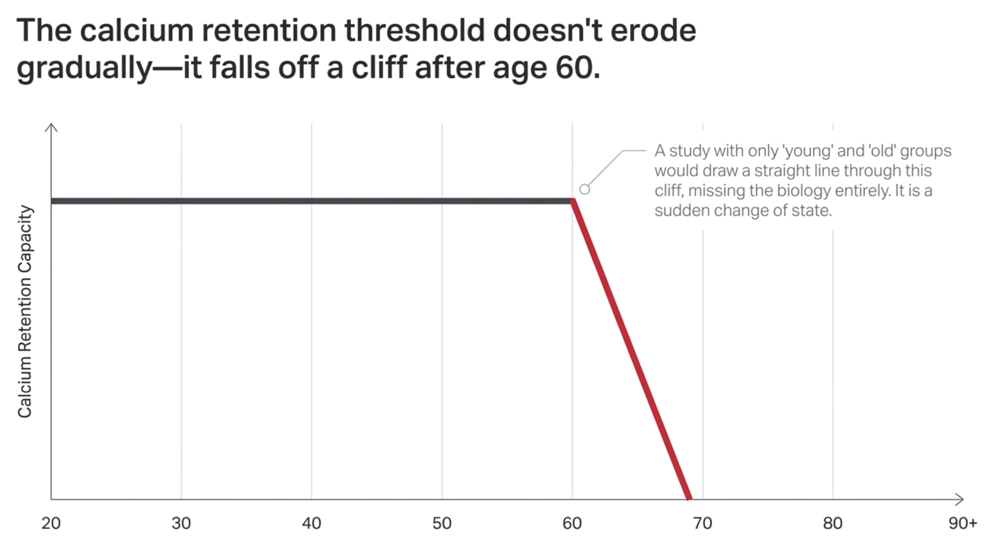
The decline is not gradual, it is a change of state after 60. Calcium retention capacity held nearly flat from the twenties through the fifties, then dropped sharply after 60. This matters because it means the risk builds in a specific decade rather than eroding evenly, and a study using only a young and an old group would have missed the shape entirely, drawing a straight line through a cliff.
When the pore opens, it links muscle aging to inflammation. A pore that opens too easily spills mitochondrial DNA into the cell, where the immune system treats it as a bacterial invader and triggers the same cGAS-STING and inflammasome alarms behind chronic age-related inflammation. This is a plausible mechanistic bridge between failing muscle and the broader "inflammaging" that accompanies aging, though the downstream steps were established in other work, not measured in these men.
This is one careful study, and its limits are real. It included only men, was cross-sectional so it cannot prove the calcium defect causes muscle loss rather than accompanying it, and drew from an unusually healthy, high-socioeconomic-status cohort. A serious alternative explanation, that age-related nerve loss drives both the muscle decline and the calcium changes, remains open, and the analyses that would help resolve it are not yet published.
The practical lesson is to measure your trajectory before decline announces itself. The defect this study identifies is invisible while it develops, accelerates in a specific decade, and leaves traces in physical function, body composition, and blood markers. Like blood pressure or cholesterol, the window to act is before symptoms appear, on the evidence of a measured number rather than a feeling. No single marker tells the story, but a panel tracked over time can show where you stand and where you are heading.
The Confound Nobody Controlled For
Ask most people who study aging why an eighty-year-old is weaker than a thirty-year-old, and you will get some version of the same answer: the mitochondria wear out. They burn less fuel, they leak more free radicals, and the muscle slowly gives up. It is one of the most durable explanations in biology, it appears on nearly every list of the hallmarks of aging, and it has guided where the field has spent its money for the better part of fifty years.
The stakes are not academic. Sarcopenia, the age-related loss of muscle mass, strength, and function, affects roughly 14 percent of people between 65 and 70 and more than half of those past 80. Somewhere over 50 million people have it now, and demographic projections put that number above 200 million within thirty years. Losing muscle is how mobility ends, how falls begin, and how independence quietly runs out.
But there is a problem with the evidence underneath the standard explanation, and it is embarrassingly simple. Older people move less. Nearly every human study of mitochondrial aging has compared a group of young adults to a group of older adults and attributed the difference to the passage of time. Those studies were also, without meaning to be, comparing an active group to a sedentary one.
That would not matter if muscle mitochondria were slow to respond. They are not. They are among the most plastic structures in the body, and they respond to load on a timescale of days. Twelve weeks of training substantially raises both mitochondrial content and respiratory capacity in older adults. Run the experiment in the other direction and the effect is just as fast: ten to fourteen days of bed rest measurably lowers mitochondrial content and respiration in older adults, while a single hour of daily exercise is enough to cancel the effect of that bed rest entirely. A mitochondrial profile is not a readout of how old someone is. It is closer to a readout of what they did last month.
The same problem contaminates the animal work used to corroborate the human findings. Aged rodents reliably show impaired mitochondrial respiration, and aged rodents also move far less than young ones in the cage. Two decades of studies may have been measuring sedentariness in a species that cannot tell us it stopped exercising.
Physical activity, in other words, is not a nuisance variable in this field. It is plausibly the variable, sitting inside every dataset that has been read as evidence for mitochondrial decline. Which raises a design question with an obvious answer and an expensive execution: to learn what aging does to mitochondria, you have to measure how much people actually move, hold it constant, and then look at what is left.
That is the study Marina Cefis, Gilles Gouspillou, and their colleagues at the Université du Québec à Montréal published in Cell Reports Medicine in early 2025. They recruited 139 men between the ages of 20 and 93, sorted them by whether they were meaningfully physically active, took muscle biopsies from all of them, and put three separate mitochondrial functions on trial at once. Two of the three turned out to be innocent.
How You Build a Study That Can Separate the Two
Three decisions made this design work, and none of them are the kind of thing that shows up in a press release.
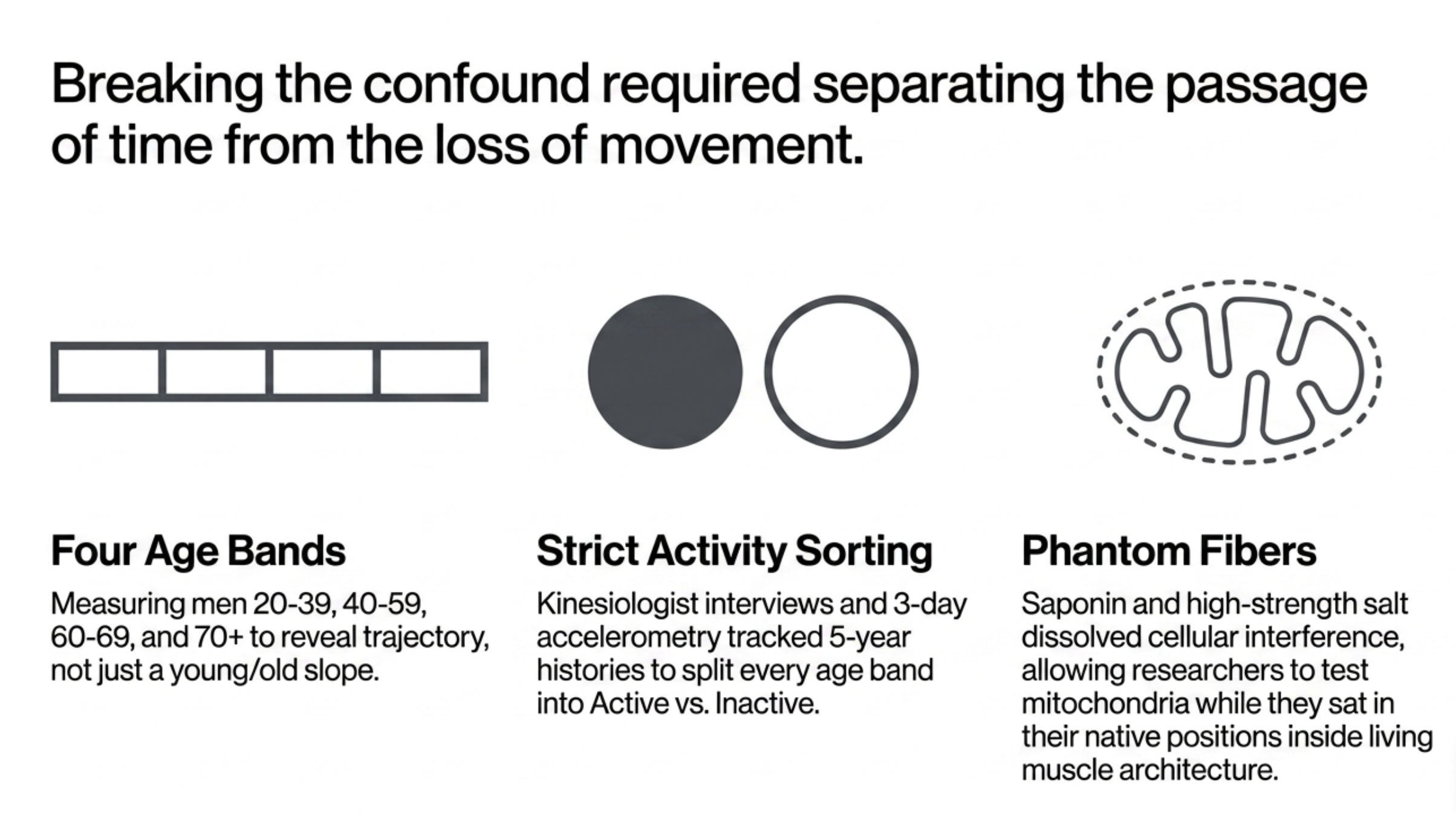
The first was refusing to study only the ends of the lifespan. Instead of a young group and an old group, the men were sorted into four bands: 20 to 39, 40 to 59, 60 to 69, and 70 and older, with active and inactive subgroups inside each. This sounds like bookkeeping. It turns out to be the difference between seeing a trajectory and seeing a slope. One of the paper's central findings is a measure that holds almost perfectly steady for forty years and then falls off sharply after 60. A study with two age groups would have drawn a straight line through that and reported a gradual decline, which is the wrong shape, and points at the wrong biology.
The second was measuring physical activity twice, using methods that fail in different directions. Every participant wore a tri-axial accelerometer for at least three days, and only days with at least 80 percent wear time counted. Every participant also sat for a structured interview with a kinesiologist, adapted from a validated activity questionnaire but modified in one important way: instead of asking about the past week, it asked what the person's activity had typically looked like over the past five years. To be classified as active, a man had to meet at least one of three thresholds: 150 minutes per week of structured moderate-to-vigorous activity, 10,000 steps per day, or an average metabolic equivalent of 1.6 or higher. Fifty-one men landed in the inactive group and 88 in the active group.
The authors are candid about where the accelerometer fails. An armband undercounts swimming, cycling, hockey, and resistance training, which means it systematically underestimates exactly the people who are most active. That limitation shapes how the data can be read, and to their credit the researchers respect it: whenever they use step count as a continuous number, they restrict the analysis to inactive men, the only group for whom the device is trustworthy.
The third decision was to measure mitochondrial function in living tissue rather than infer it from a blood marker or an imaging proxy. Each man underwent a biopsy of the vastus lateralis, the outer quadriceps, taken with a suction-modified needle under local anesthetic. What happened next is where the study earns its findings.
Muscle fibers are wrapped in a plasma membrane that keeps experimental substrates out. The standard workaround is to grind up the tissue and spin the mitochondria out into a pellet, but mitochondria pulled out of a cell behave differently from mitochondria left in one, and specifically they exaggerate age-related impairment. The alternative is a chemical trick. Saponin binds cholesterol, and the outer membrane of a muscle fiber is rich in cholesterol while mitochondrial membranes contain almost none. Applied at low concentration, it perforates the cell surface while leaving the mitochondria untouched, still sitting in their native positions inside the fiber's architecture. It is the difference between taking the roof off a factory to watch the machines run and hauling the machines out into a parking lot to test them.
Even that is not enough to measure calcium. Actin and myosin, the proteins of contraction, bind calcium with such affinity that any calcium added to a permeabilized fiber is captured by the contractile machinery before the mitochondria ever see it, like trying to measure how much water a sponge absorbs while it sits buried in paper towels. So the team went further and made what the field calls phantom fibers. A high-strength salt buffer dissolves the myosin out of the fiber while leaving mitochondrial function intact. What remains is a ghost of a muscle fiber with its mitochondria still in place and nothing left to compete with them for calcium. Only then can you flood the preparation with calcium and watch, using a fluorescent indicator, exactly how much the mitochondria absorb before something gives way.
Respiration and free-radical emission were recorded at the same time in a high-resolution respirometer at body temperature, with the electron transport chain fed different fuels in sequence to interrogate specific entry points. Fiber typing was done on more than 200 individually traced fibers per participant, by an experimenter blinded to each man's age and activity status.
What this design can establish is what differs between groups. What it cannot establish is what causes what. This is a cross-sectional study, so every finding is a snapshot of different men at different ages rather than a trajectory followed through time. Activity status was self-selected, not assigned, so the active men differ from the inactive men in ways beyond exercise. Those constraints matter most at the end, and they do not weaken what comes next, because what comes next is mostly a set of things that did not happen.
Suspect One: The Failing Engine
Before anything can be explained, the thing being explained has to be real, and in these 139 men it was. Six-minute walk distance, step-test performance, sit-to-stand repetitions, and timed up-and-go all declined significantly with age. Maximal knee-extension strength fell, and lower-limb power fell faster. Thigh muscle cross-sectional area shrank while the fat threaded between the muscles increased. This is the phenotype the mitochondrial theory was invented to account for, and it showed up exactly on schedule.
Physical activity blunted it without stopping it. Active men outperformed inactive men on every functional test at nearly every age, and the protection widened with age rather than holding constant. Activity does not flatten the curve of functional decline. It tilts it.
Two details in this opening picture deserve more attention than they usually get, because both foreshadow what the mitochondrial data will show. First, active men were stronger and more powerful only when strength was expressed relative to body mass. Their absolute strength, and their strength per unit of thigh muscle, showed no significant activity advantage. The active men were not producing more force from each unit of muscle. They were carrying less mass with the muscle they had. Second, and more striking, physical activity had no effect at all on muscle cross-sectional area or thigh lean mass. The active men lost muscle on the same schedule as everyone else. What activity protected was function, body composition, and the fat infiltrating the muscle, not muscle size.
Hold onto that. It is the first hint that something in the aging process is running underneath exercise rather than through it.
Now the engine. If mitochondrial decline drives muscle aging, maximal respiratory capacity should fall with age. It did not. Maximal respiration supported by the standard fuels showed no effect of age across the four bands. Treated as a continuous variable across the whole cohort, the relationship did not reach significance, and within the active men alone it was effectively nonexistent. A man in his ninth decade who had kept moving had muscle mitochondria that respired like a man in his twenties.
Fat oxidation told the same story. Respiration supported by fatty acids, the entry point for burning fat, was unchanged across the lifespan. This matters more than the headline, because fat is the dominant fuel at rest and during the low-intensity movement that fills an ordinary day. If aging muscle were failing to burn fat, it would fail during the very activities that constitute daily living. It was not.
What did change was activity status, and it changed everything. Active men had higher maximal respiration, higher fat-supported respiration, and higher citrate synthase activity, a standard proxy for how much mitochondrial material a muscle contains. The effect was large and consistent across age bands.
Then comes the result that reframes the section. When respiration was normalized to citrate synthase activity, to ask how well each unit of mitochondria performs rather than how much total capacity the muscle has, every difference vanished. Not just the age trend. The activity advantage disappeared too. Exercise does not build better mitochondria. It builds more of them. This aligns with recent work showing that respiration per unit of mitochondrial membrane is indistinguishable between untrained people, recreational exercisers, and elite runners. The machines are identical. Trained muscle simply runs a larger factory.
There is a residual signal worth reporting honestly. Among inactive men, respiration did trend downward with age, and mitochondrial content trended down alongside it, though the trend fell short of significance. But within that same inactive group, daily step count positively predicted both respiration and mitochondrial content, and step count itself declined with age. The trend is a movement effect wearing an aging costume, which is precisely the confound the study was built to expose.
One more finding cuts against a specific version of the mitochondrial theory. The idea that aging mitochondria become mildly uncoupled, wasting fuel as heat instead of capturing it as usable energy, predicts falling coupling efficiency. The study found the opposite: coupling efficiency was preserved, and by one measure slightly improved, with age.
None of this means mitochondrial capacity is irrelevant. Across the pooled cohort, respiration correlated positively with muscle size, strength, power, and physical performance, replicating earlier findings. Men with better mitochondria were doing better. The claim is narrower and more specific: mitochondrial respiratory capacity does not decline as a function of age itself, so whatever is causing muscle to fail with age, it is not a fuel-burning problem.
One suspect down. The second has a longer history and a larger following.
Suspect Two: The Leaking Engine
Denham Harman proposed in 1956 that aging is the accumulated damage of free radicals, and sixteen years later he named the mitochondrion as the primary source. It became the most influential idea in the biology of aging, it built an industry, and it has been failing its tests for two decades. This study is one of the harder tests it has faced in human muscle.
Hydrogen peroxide emission was measured in the same fiber bundles, at the same time, as respiration, using a fluorescent probe that reports free-radical release in real time. Peroxide is the standard readout for mitochondrial free-radical production. The team measured it under several different fuels and under a maximal condition designed to force electrons to leak.
Aging did nothing in any of them. Across four decades of separation and every substrate condition the team could construct, mitochondrial free-radical output was flat. Older mitochondria did not leak more than younger ones.
The activity result is the one that turns the section. Active men emitted more hydrogen peroxide than inactive men, not less, in every condition where activity mattered at all. The men who walked farther, stood up faster, and carried less fat between their muscle fibers were the men whose mitochondria released the most peroxide. Once you know that exercise builds more mitochondria rather than better ones, this stops being a paradox. More factories, more exhaust.
That interpretation is confirmed by the normalization. When peroxide emission was expressed as a fraction of oxygen consumed, revealing whether each mitochondrion was intrinsically leakier, there was no effect of age or activity. Nobody's mitochondria were intrinsically leakier. When emission was instead normalized to mitochondrial content, group differences again disappeared, with one partial exception the authors are careful to state: among inactive men only, and only under one fuel condition, content-normalized emission did rise with age. It did not hold under the other conditions. It is a thread rather than a finding, and it belongs to the same inactive group whose respiration trend traced back to declining step counts.
The strongest evidence against the free-radical account is the absence of any dose-response relationship with the outcome. If free radicals were eroding muscle, the men producing the most should be the smallest and weakest. The study found no negative association between peroxide emission and muscle mass, strength, or physical performance anywhere in the dataset. The relationship, if anything, ran the other way, because the highest emitters were the fittest men in the cohort.
This has direct implications for a category of supplement, and they are worth spelling out.
The first problem is the premise. Mitochondria-targeted antioxidants are sold on the assumption that aging muscle produces excess mitochondrial free radicals that need neutralizing. In healthy human muscle spanning seven decades, that excess does not appear. There is nothing at the target.
The second problem is that the interventions have already been tested against the outcome that matters, and they failed. Long-term administration of MitoQ, a mitochondria-targeted form of coenzyme Q10, did not reduce age-related oxidative damage or rescue the loss of muscle mass and function in rodents. A related compound, SS-31, produced the same null result. The cleanest test is genetic: mice engineered to lack manganese superoxide dismutase, the primary antioxidant enzyme inside the mitochondrial matrix, live with elevated oxidative stress and impaired mitochondrial function and still do not develop muscle atrophy with age. If maximal, lifelong, unopposed mitochondrial free-radical exposure does not cause muscle wasting, then blocking a normal amount of it is unlikely to prevent muscle wasting.
The third problem is what the fitness data imply about the signal itself. There is a separate and not fully settled literature suggesting that high-dose vitamin C and E taken around training can blunt some of the adaptations exercise is meant to produce, consistent with free radicals functioning as a signal the muscle uses rather than only as a toxin it suffers. That work sits outside this study and should not be leaned on heavily, but it points in the same direction as the observation that the healthiest men here were the highest emitters.
The scope of this conclusion needs stating as plainly as the conclusion itself. This is a finding about mitochondrial free-radical production in skeletal muscle, in healthy, community-dwelling men. It is not a verdict on antioxidants for cardiovascular, neurological, or eye conditions, where the biology and the evidence are different. It says nothing about pathological aging or muscle under conditions of illness. And it does not mean oxidative stress is irrelevant to human biology. It means that in this tissue, in this population, mitochondrial free-radical overproduction is not what makes an old muscle weak.
Two suspects have now been eliminated by the same logic: the function was measured, it did not change with age, and it did not track the outcome. The third suspect breaks that pattern in both directions.
Suspect Three: The Trigger That Fires Too Easily
Mitochondria have a second job that gets almost no attention outside cell biology. Besides making ATP, they absorb calcium. A muscle fiber floods itself with calcium every time it contracts, then pumps it back into storage, and the mitochondria sitting inside that flood act as buffers, taking up the calcium that escapes and holding it. The same proton gradient that drives ATP synthesis also drives calcium into the mitochondrion, so the same machinery does both jobs.
Every buffer has a limit, and this one fails in a specific and violent way. When the calcium inside a mitochondrion exceeds what it can hold, a large channel called the mitochondrial permeability transition pore snaps open in the inner membrane. The membrane potential collapses. The organelle swells, dumps its calcium back into the cell, and spills its contents, including proteins that are dangerous outside the mitochondrion, into the surrounding cytoplasm. The pore is not a slow leak. It is a threshold event, and crossing it is the mitochondrial equivalent of a decision to stop.
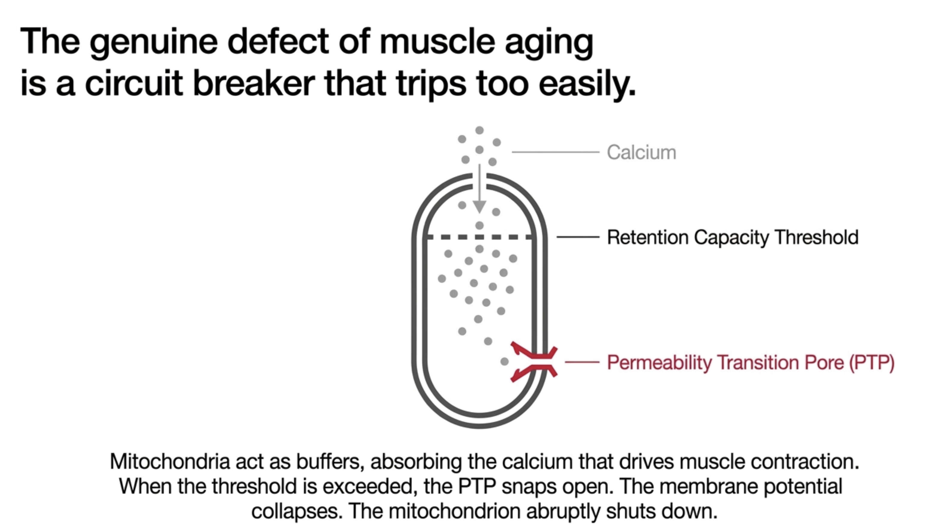
The assay measures where that threshold sits. Phantom fibers were flooded with calcium while a fluorescent indicator tracked how much disappeared into the mitochondria. The total absorbed before the pore blew open is the calcium retention capacity. How long it took is the time to pore opening. Together they describe how much stress a mitochondrion will tolerate before it triggers.
Both fell with age. Calcium retention capacity declined significantly across the four age bands, with the 70-plus group differing from both the youngest men and the 60-to-69 group. Time to pore opening declined as well. After two mechanisms that refused to change with age, this one changed.
Physical activity did nothing. Not partial protection, not an attenuated slope, nothing. The active men in this cohort walked farther, stood faster, carried less fat between their muscles, had better insulin sensitivity, and had substantially more mitochondrial machinery. Their mitochondria triggered just as early as everyone else's.
The shape of the decline matters as much as its existence, and it is the reason the four age bands were worth the expense. Calcium retention capacity is essentially flat from 20 to 60. Then it falls sharply. A study comparing thirty-year-olds to seventy-five-year-olds would have found a difference and drawn a gradual line between them, describing a slow erosion that never actually happened. What the data show instead is a stable system that changes state somewhere in the sixth decade.
Retention capacity also tracked the outcomes that matter. It correlated with thigh lean mass, with knee-extension strength, with six-minute walk distance, and with step-test performance. These correlations need to be reported with their real strength, because they are weak. The strongest of them explains only a small fraction of the variance between men, and two of them sit close enough to the threshold of significance that either could fail to replicate in another cohort. No single one of these would justify a paragraph on its own.
The argument is not any one correlation. It is that four independent outcomes, measuring different things through different instruments, all point the same direction, and that the mechanistic story predicts exactly that direction. Weak and consistent is a different kind of evidence from weak and scattered. It is still not strong evidence, and the honest reader should hold it as suggestive rather than settled.
Two other cohorts have found the same decline. Calcium retention capacity was reduced in recreationally active older adults in earlier work from this group, and reduced again in master athletes, men who had trained seriously for decades. Whatever this is, it is not rescued by doing more of what already works.
Now put the pieces together. Physical activity protected function, body composition, insulin sensitivity, intermuscular fat, and mitochondrial content. It did not protect muscle cross-sectional area, and it did not protect strength normalized to the muscle producing it. The active men got weaker and smaller on roughly the same schedule as the inactive men, while performing far better on every functional test. Something is driving that residual loss, and it is running underneath exercise rather than through it. Of everything measured in these 139 men, mitochondrial calcium handling is the only variable that behaves the same way: declining with age, indifferent to training, and tracking the outcomes that were lost.
That is a strong fit, and it is not proof. Because this is a cross-sectional study, it cannot establish that failing calcium buffering causes muscle loss rather than simply accompanying it. There is at least one serious alternative worth naming. Motor neurons die with age, and muscle fibers that lose their nerve supply show mitochondrial abnormalities. The master-athlete study that found reduced retention capacity attributed it to this denervation rather than to aging of the mitochondria themselves. If the loss of motor neurons is upstream of both the muscle loss and the calcium phenotype, then calcium handling is a symptom wearing the clothes of a cause. The same group collected these biopsies with a method designed to enrich for neuromuscular junctions, and those analyses are still unpublished, which means the question is open, and the people best positioned to answer it are already working on it.
What the study does establish is narrower and still substantial. In healthy men across seven decades, the mitochondrial function that changes with age is not energy production and not free-radical output. It is the threshold at which the mitochondrion pulls its own trigger. And unlike everything else measured here, that threshold does not respond to the one intervention we know works.
Before following the pore downstream into the muscle, there is one more finding to account for, because it suggests this may leave a trace in the blood.
The Signal That Shows Up in Blood
Muscle biopsies do not scale. If mitochondrial calcium handling matters, the practical question is whether anything about it can be seen without a needle in the quadriceps, and the study offers a partial answer.
Growth differentiation factor 15, or GDF15, is a protein that cells release when they are under stress, and mitochondria under stress release it especially reliably, which has earned it the nickname of mitokine. In this cohort, plasma GDF15 tracked age closely, one of the strongest relationships in the entire dataset. The rise was not linear. Concentrations climbed steeply, and the climb accelerated after 60, at the same point where calcium retention capacity falls off its plateau. Physical activity did not significantly lower it.
GDF15 also tracked mitochondrial function in the expected directions. Men with higher levels had lower respiratory capacity, an association that reached significance only among the inactive. Men with higher levels also had lower calcium retention capacity, meaning mitochondria that trigger sooner. That particular correlation was weak, and it carries a caveat the authors disclose: the GDF15 correlations were tested with a more permissive statistical standard than the rest of the paper, which means they warrant more caution, not less.
The temptation here is to treat GDF15 as a blood test for mitochondrial aging, and that temptation should be resisted. GDF15 rises in pregnancy, in kidney and cardiovascular disease, in cancer, in smokers, and, notably for anyone in the longevity world, in people taking metformin. It is a general stress signal, not a calcium assay, and a single elevated value tells you that something is straining without telling you what. On its own, it cannot isolate the mitochondrial process this study describes.
What GDF15 contributes here is narrower and still useful: the muscle-level finding leaves a systemic footprint, and that footprint bends upward on the same timeline. Two independent measures, one requiring a biopsy and one requiring only a blood draw, change state in the same decade. That convergence is the point. No single blood marker will ever capture something as specific as a mitochondrial calcium threshold, but a marker that moves with the underlying biology, read alongside other measures and tracked over time rather than in isolation, is exactly the kind of signal that turns an invisible process into something you can watch. The value is not in one number on one day. It is in the trajectory, and in reading it in context.
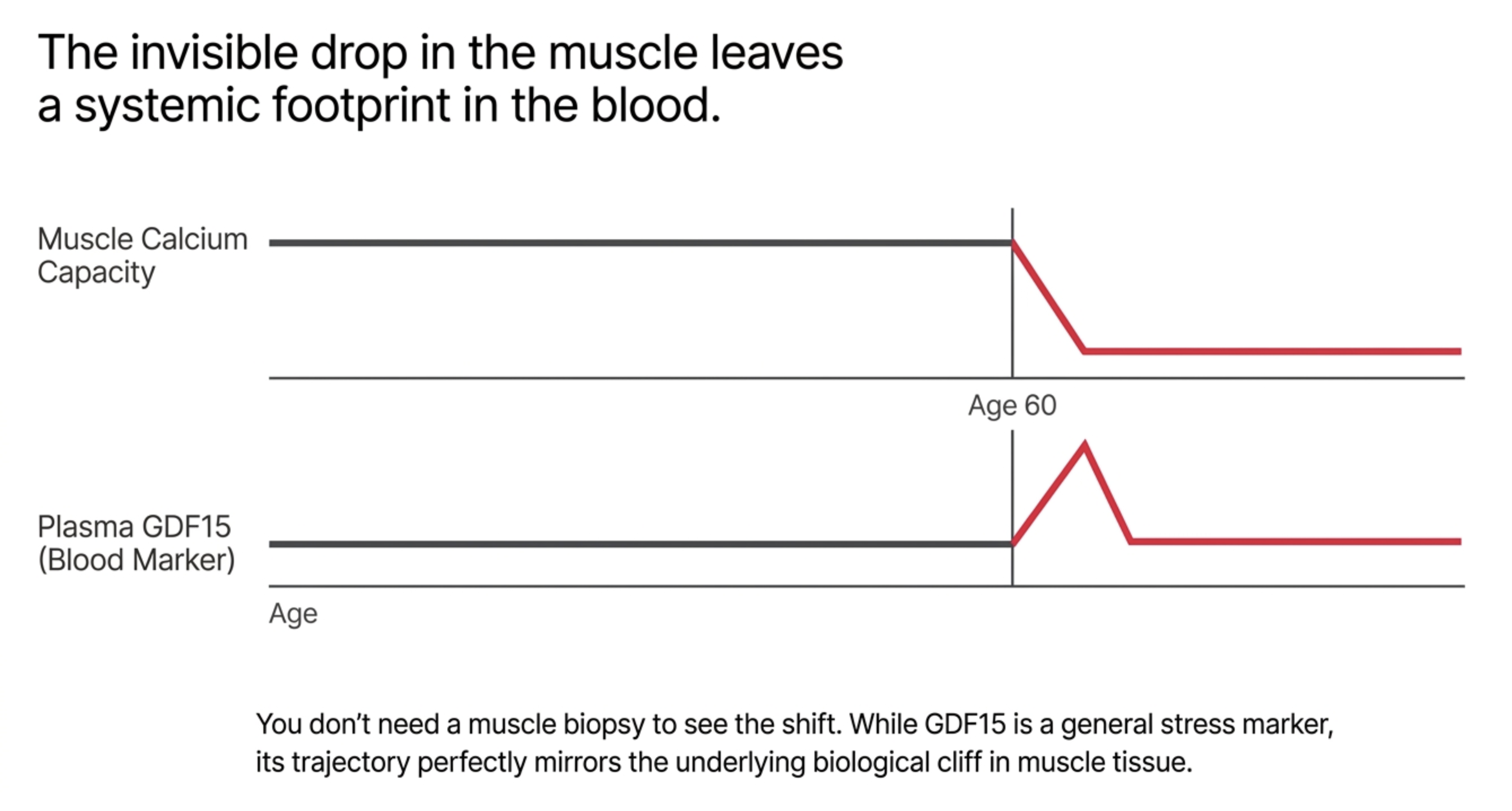
That decade, the one where both the muscle measure and the blood measure change state, is where the mechanism becomes urgent, because a pore that opens too easily does not simply fail to buffer calcium. It sets off a sequence.
What Happens When the Pore Opens
Think of the permeability transition pore as a circuit breaker. The wiring in an aging muscle is not obviously damaged, and the load it carries has not changed much. What changes is the current at which the breaker trips. Set it low enough and the system starts shutting itself down during ordinary use, and each trip costs something that does not get fully rebuilt.
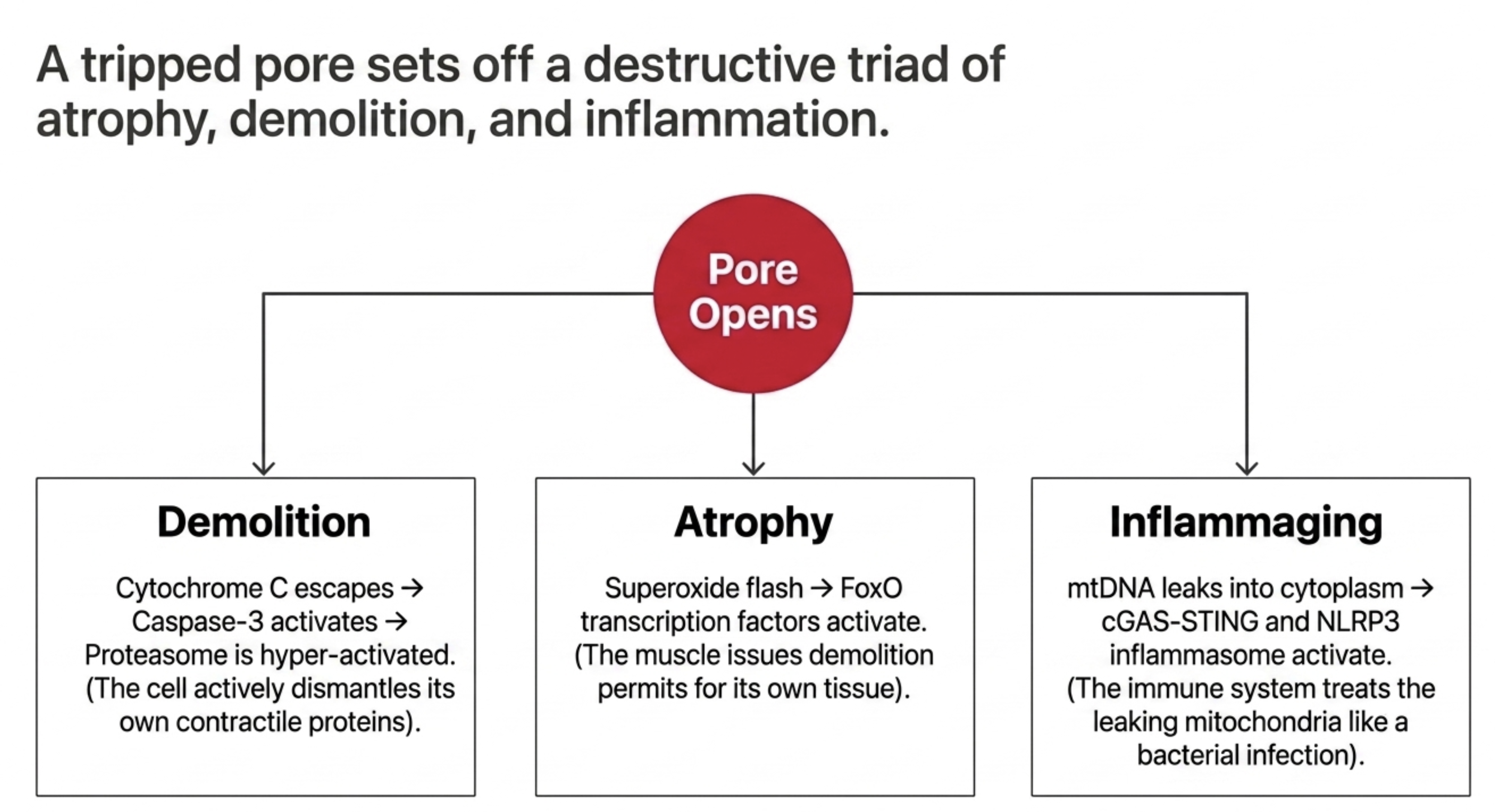
Several consequences follow from pore opening, and they are not competing explanations. They run in parallel, from a single event.
The first is protein breakdown. When the pore opens, a protein called cytochrome c escapes into the cytoplasm, where it triggers the activation of caspase-3. Caspase-3 is known for its role in programmed cell death, but it does something more specific in muscle: it cleaves part of the proteasome, the cell's protein-disposal machinery, in a way that makes the proteasome more active. A single pore event therefore hands the fiber a sharpened tool for dismantling its own contractile proteins.
The second is the atrophy program itself. Pore opening produces a brief, intense burst of free radicals, visible in single mitochondria as what researchers named a superoxide flash. Free radicals in that concentrated, transient form activate a family of transcription factors called FoxO, and FoxO activation switches on the two molecular tags that mark muscle protein for destruction. This is the muscle equivalent of issuing demolition permits.
That paragraph appears to contradict everything established two sections ago, and the resolution matters. The section on free radicals measured steady-state emission from the electron transport chain, averaged across a fiber bundle over minutes, under controlled conditions. What follows pore opening is different in quantity, location, and duration: a transient flash, confined to individual mitochondria, over seconds. An assay built to capture bulk, background emission would not detect intermittent flashes in a small subset of organelles, and the two measurements are not in competition. Free radicals in this model are not a chronic background insult that gradually corrodes the muscle. They are a downstream signal, released by a trigger that fires too easily. Which is also why flooding the tissue with antioxidants failed. You cannot fix a breaker by insulating the sparks.
The third consequence connects muscle aging to inflammation, and it links this study directly to biology we have covered before. Mitochondria carry their own DNA, a relic of their ancient bacterial ancestry, and the immune system still treats that DNA as foreign when it appears outside the organelle. When the pore opens, fragments of mitochondrial DNA escape into the cell, where they activate the same internal alarm systems, the cGAS-STING pathway and the NLRP3 inflammasome, that we described in an earlier article on mitochondria and inflammation. Those sensors respond as though they had detected an infection, igniting inflammation, and that inflammation has in turn been linked to the very same protein-tagging machinery that drives muscle atrophy. A leaky pore does not just weaken the fiber locally. It supplies the innate immune system with a reason to treat the muscle as damaged tissue, which is a plausible mechanistic bridge between muscle aging and the chronic, low-grade inflammation that pervades the aging body. The calcium threshold falling in these men is, in this light, one upstream tap feeding the inflammaging described in that earlier piece.
There is a fourth consideration that is less a consequence than a compounding factor, and it may explain the one thing the calcium finding otherwise leaves unexplained. Aging muscle does not only have a smaller mitochondrial calcium buffer. It also has more calcium to buffer. Resting calcium in the fiber is elevated with age, the channel that releases calcium during contraction becomes oxidized and leaks, and the pump that returns calcium to storage works less efficiently. A smaller reservoir facing a heavier load is worse than either problem alone.
This is where exercise may still be doing something, and it resolves the tension at the center of the whole study. Resistance training has been shown to partially restore the muscle's calcium-storage pump, which would lower the ambient calcium load without changing the mitochondrial threshold at all. If that is what happens, then physical activity is not protecting the buffer; it is reducing what the buffer has to handle. That would account for the pattern the study found: functional protection that is real and substantial, sitting alongside a calcium retention capacity that declines on schedule regardless. Exercise does not raise the trigger threshold. It may simply keep the muscle from pushing against it as hard.
One caution about this entire section. Cefis and colleagues measured where the threshold sits. They did not measure the downstream steps, the caspase activation, the protein-tagging, the inflammatory signaling, in these men. The chain that follows the pore is assembled from rodent work, cell biology, and earlier human studies, and it is well supported at each link, but the links were established elsewhere. What this study contributes is the observation that the initiating event becomes more likely with age in humans, and that the change is untouched by training. The sequence that follows is inference, built on solid ground, and still inference.
Which leaves the question of what to do with it.
What This Changes, and What It Does Not
It is worth separating what this study settles, what it opens up, and what it merely licenses people to speculate about, because those three categories tend to collapse into one another the moment a finding reaches an audience looking for something to do.
Start with what is established, because it is the most important part and the easiest to lose in the excitement over calcium. Exercise remains the most effective intervention available for aging muscle, and nothing in this paper qualifies that. The active men walked farther, stood up faster, moved through the timed tests quicker, and carried more strength and power per unit of body mass at every age. They had less fat overall and less fat infiltrating the muscle, lower fasting glucose and insulin, better insulin sensitivity, and lower triglycerides. They had more mitochondrial machinery and greater respiratory capacity, including the capacity to burn fat, into their ninth decade. A ninety-year-old who kept moving had mitochondria that respired like a young man's.
The reframe this paper supports is not that exercise matters less. It is that exercise is the floor rather than the ceiling. It protects nearly everything, and the few things it does not protect are now visible: muscle size, strength normalized to the muscle producing it, and the mitochondrial calcium threshold. That gap is not an argument for training less. It is an argument that training alone will not close it. And it fits a theme running through our recent coverage: a companion study on anabolic resistance found that exercise re-sensitizes aging muscle to dietary protein, and this one finds that exercise preserves mitochondrial number and physical function. Different mechanisms, converging on the same intervention, with the same honest asterisk that it is powerful but not total.
Then there is what is emerging. If the initiating event in muscle aging is a permeability transition pore that opens too easily, then the target is anything that raises that threshold or lessens the calcium load pressing against it. Several approaches are conceivable: stabilizing the regulators that govern how readily the pore opens, calming the leaky channel that raises resting calcium, or modulating the route through which calcium enters the mitochondrion in the first place. None of this has been tested against muscle aging in humans, and the compounds that might do it cleanly do not yet exist in usable form.
There is one compound worth watching precisely because it already failed, and the reason it failed may have been the wrong test rather than the wrong drug. SS-31, also called elamipretide, entered the aging field labeled as a mitochondria-targeted antioxidant and was tested against that hypothesis in rodent muscle, where it did not rescue mass or function. But scavenging free radicals is not really what the compound does. It binds cardiolipin, a distinctive fat found almost only in the inner mitochondrial membrane, and stabilizes the membrane architecture that cardiolipin organizes, architecture that is itself involved in permeability transition. Under the framework this paper proposes, the drug may have been given the right mechanism and the wrong rationale, then judged against an endpoint chosen by the theory this study is dismantling. That is a hypothesis to hold loosely, not a recommendation. The rodent result was null regardless of the reasoning, the human data for this compound come from rare genetic mitochondrial diseases whose biology does not transfer to normal aging, and nothing here is a reason for anyone to seek out peptides. It is a reason to watch one specific question: whether a compound aimed at membrane stability behaves differently when it is finally tested against the endpoint this paper implicates.
And then there is what remains genuinely speculative: whether the sharp decline after 60 is a true change of state or an artifact of splitting a continuous process into bands; whether the calcium defect causes muscle loss or merely accompanies it, with denervation the most credible upstream alternative and the deciding analyses still unpublished; and whether any of this holds in women, whose neuromuscular aging follows a less linear course and who were excluded from this study entirely. One more honesty check on a claim made earlier: calcium handling is the only variable measured here that fits the pattern of the residual loss, which is not the same as the only variable that could fit it. The study examined three mitochondrial functions. Muscle aging is not obliged to be a mitochondrial problem at all.
The limits of the cohort bound all of this. Only men were enrolled, a deliberate choice justified by the more linear course of male neuromuscular aging, but a real constraint on who these findings describe. Nearly all of the participants were of high socioeconomic status, and the cohort was overwhelmingly white. Every man was community-dwelling and in good general health, which means the inactive group was not a sedentary-and-sick group; most of them, including the inactive, would meet a reasonable definition of successful aging. The mitochondrial changes seen in frail or chronically ill older adults could look quite different. And the design is cross-sectional, so no trajectory was ever actually observed; the sharp post-60 drop is inferred from different men measured once, not the same men followed down the slope.
What survives all of that is a single, sturdy observation. Across seven decades of human muscle, what aged was not the capacity to make energy and not the tendency to leak free radicals. What aged was the point at which the mitochondrion pulls its own trigger. And that brings the practical question back to the one thing a person can act on today, which is not a drug and not even, entirely, exercise. It is knowing where they stand.
Because the decline this study describes is not uniform. It runs on different timelines in different people, it accelerates in a specific decade, and it leaves traces that can be measured, in physical function, in body composition, and in blood markers that bend upward as the underlying biology shifts. The lesson is not to wait for weakness to announce itself, because by the time it does, the change of state has already happened. The lesson is to measure the trajectory of your own aging while it is still quiet, and to track it over time rather than guess. That is the premise Healthspan's BioAge+ is built on: that the course of your aging is measurable, that no single number tells the story but a panel read over time can, and that measuring it is the first move toward changing what can still be changed, and watching what cannot.
The Decade Before
For seventy years the field has been trying to make old mitochondria burn better and leak less. It funded the antioxidant industry, it shaped the design of hundreds of studies, and it produced a picture of aging muscle as a worn-out engine losing compression. The picture was intuitive, it fit the free-radical theory, and in healthy human muscle it appears to be wrong. These mitochondria burn fine. They do not leak more. A man in his nineties who kept walking had oxidative machinery that would not embarrass a graduate student.
What changed across those seven decades was not the engine's output. It was the threshold at which the mitochondrion shuts itself down, and everything that follows from crossing it: the disposal machinery sharpened, the atrophy program licensed, mitochondrial DNA spilled where the immune system can find it. The failure is not a loss of power. It is a trigger that has become too easy to pull.
That distinction is not academic, because the two framings point at completely different interventions. If the problem is a weak engine, you build more capacity, and exercise does that better than anything in a bottle. If the problem is a sensitive trigger, capacity is beside the point, which is exactly what these 139 men demonstrated. The active ones had more mitochondria, more respiratory capacity, better function on every test, and thresholds that had fallen just as far as everyone else's. They were doing the right thing. It simply was not addressing this.
The timing is the part worth sitting with. Calcium retention capacity in this cohort held nearly flat from the twenties through the fifties, then dropped after 60. The blood marker that tracks mitochondrial stress bent upward on the same schedule. Whatever is happening looks like a change of state rather than a slow erosion, and it happens in a decade most people spend feeling fine.
Which means the window for anything that eventually targets this is not the decade after the decline. It is the decade before. That is an uncomfortable place to intervene, because the people in it have no symptoms, no complaints, and no reason to think anything is coming. It is also where nearly every successful preventive intervention in medicine has had to operate. Blood pressure, cholesterol, bone density: in each case the win came from acting before the damage announced itself, on the evidence of a number rather than a symptom.
None of this is settled. The correlations are modest, the design cannot establish cause, the cohort was narrow, and the compounds that might one day raise a mitochondrial threshold in a living person do not yet exist in usable form. What has changed is the question being asked. For most of a century the field asked why old muscle runs out of energy. The better question, on this evidence, is why old muscle becomes so willing to take itself apart, and the answer points less toward burning brighter than toward holding the trigger a little more steadily, in the years before it starts to slip.
Related studies